
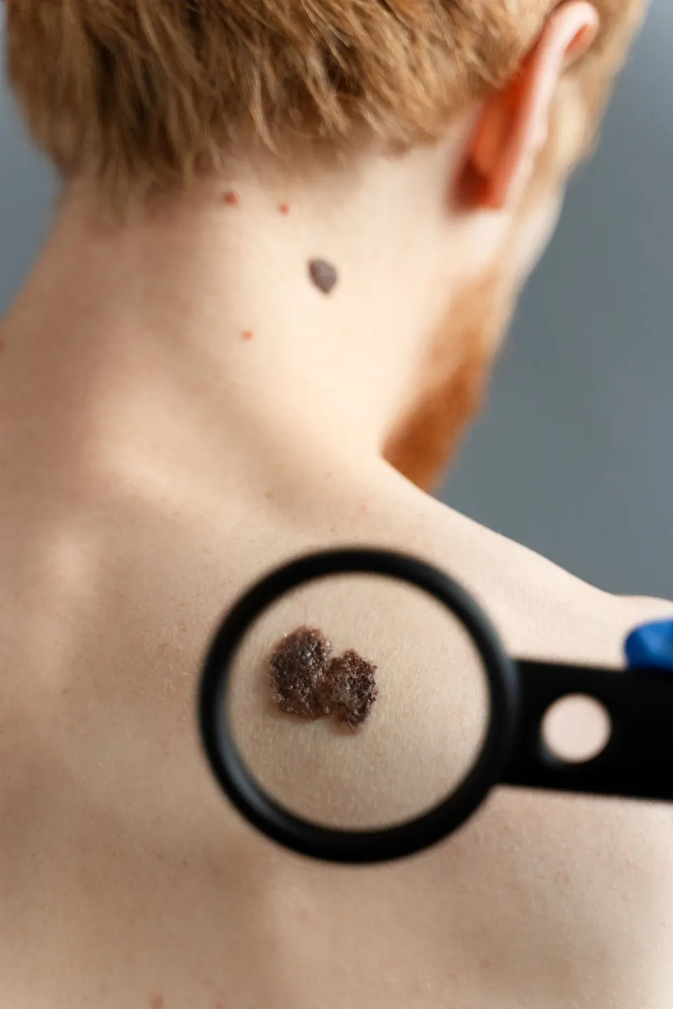
Medical content by Elias Tsiolis, Director Surgeon · Last updated: 11 May 2026
Skin Cancer
Everything You Need to Know About Skin Cancer
Types
- Basal cell carcinoma (BCC) – the most common; does not spread to other organs.
- Squamous cell carcinoma (SCC) – more aggressive; needs prompt removal.
- Melanoma – the most dangerous form; it can spread (metastasize).
Symptoms
- A new lesion that changes shape, color or size
- Bleeding or a sore that does not heal
- Irregular borders or colors
Diagnosis
Diagnosis is made by clinical examination and, in some cases, a biopsy of the lesion.
Treatment
Surgical Treatment
Treatment is surgical, with complete excision of the lesion with clear margins. Early removal leads to a full cure in most cases.
Frequently Asked Questions
Is skin cancer dangerous?
Yes, especially melanoma, but it can be cured if diagnosed early.
How often should I be checked?
Every year, especially if you have many moles or a history of sun exposure.
Is removal painful?
No, it is done under local anesthesia and leaves a minimal mark.
Moles (Nevi)
Moles are clusters of pigment cells (melanocytes) in the skin. Most are benign and cause no problems, but some can develop into melanoma, the most dangerous type of skin cancer.
Regular monitoring and, when needed, removal of suspicious moles is the best way to prevent it.
Everything You Need to Know About Moles
Types of Moles
- Congenital moles (present from birth)
- Acquired moles (develop over the years)
- Dysplastic moles – show atypical features and need monitoring or, more often, removal
When a mole needs checking
We use the ABCDE rule:
- A – Asymmetry
- B – Border (irregular or blurred)
- C – Color (several colors)
- D – Diameter (> 6 mm)
- E – Evolution (change in shape/size/color)
If a mole changes or bleeds, it needs to be examined promptly.
Diagnosis
- Dermoscopy – a detailed examination with a special lens
- Digital mole mapping – monitoring with photographic records
- Biopsy – for suspicious lesions
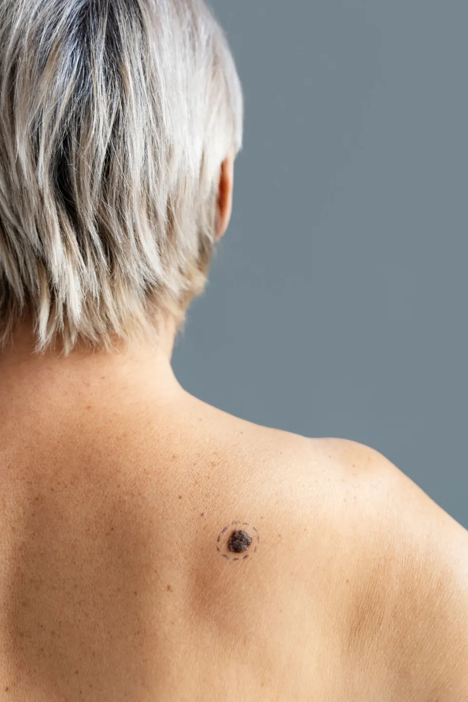
Treatment
Removal is a simple, painless procedure done under local anesthesia.
Surgical Treatment
Methods:
- Surgical excision (with stitches – for suspicious moles)
- Laser or radiofrequency (for benign moles in cosmetically sensitive areas)
- Specimens are always sent for histological examination.
Frequently Asked Questions
Will there be a scar?
Small and discreet, especially with laser or radiofrequency techniques.
When should I have my moles checked?
Once a year, or sooner if you notice a change.
Is it dangerous to have them removed?
No, as long as the removal is done properly and the specimen is sent for histology.